
Translate this page into:
Assessment of gallbladder stone – A geological approach through cutting-edge field emission scanning electron microscope and energy-dispersive X-ray fluorescence spectroscopy with anti-cancerous properties examined for hep G2 (liver) cancer cell lines with validation through reactive oxygen species anti-oxidant analysis

*Corresponding author: Yamini Malhotra, Department of Earth Sciences, Annamalai University, Chidambaram, Tamil Nadu, India. yaminipriya2008@gmail.com
-
Received: ,
Accepted: ,
How to cite this article: Malhotra Y, Subramanian SR, Vennila L, Mukesh MV, Kumar NS. Assessment of gallbladder stone – A geological approach through cutting edge field emission scanning electron microscope and energy-dispersive X-ray fluorescence spectroscopy with anti-cancerous properties examined for hep G2 (liver) cancer cell lines with validation through reactive oxygen species anti-oxidant analysis. Am J Biopharm Pharm Sci. 2024;4:4. doi: 10.25259/AJBPS_3_2024
Abstract
Objectives:
Geology, traditionally focused on the study of Earth, ocean, and planetary rocks, extends to the examination of stones formed within the human body, such as those found in the bladder or kidneys. This research specifically targets the classification and elemental composition of gallbladder stones, with a concentrated analysis on the anionic and cationic constituents. The study employs a combination of advanced imaging and spectroscopic techniques to delve into the intricate details of these stones and evaluates their potential medical applications, particularly their anti-cancer properties.
Material and Methods:
The methodology involved in this research is multifaceted, incorporating several state-ofthe-art techniques. Field emission scanning electron microscopy (FESEM) was utilized to capture high-resolution images of the gallbladder stone samples, providing a detailed look at their surface morphology. Complementing this imaging technique, energy-dispersive X-ray analysis (EDX) was employed to determine the elemental composition of the samples. Additionally, energy-dispersive X-ray fluorescence (EDXRF) spectroscopy was conducted, both with and without chromium coating, to further analyze the elemental makeup of the stones.
Results:
The results from these techniques revealed a comprehensive profile of the elemental composition of gallbladder stones. FESEM provided detailed images, allowing for a thorough examination of the stone’s microstructure. EDX analysis contributed to the identification of various elements present in the samples, highlighting the predominant anions and cations. EDXRF spectroscopy, with its high sensitivity and accuracy, corroborated these findings, ensuring a robust and precise determination of the elemental constituents.Beyond the geological analysis, the study explored the potential biomedical applications of gallbladder stones. Samples were tested for their anti-cancer properties using the MTT assay on Hep G2 liver cancer cells. The MTT assay is a colorimetric assay that measures the metabolic activity of cells, providing an indication of cell viability and proliferation. The gallbladder stones exhibited significant anti-cancerous properties, with an inhibitory concentration (IC50) value of 70.60, indicating their efficacy in inhibiting the growth of liver cancer cells. To further validate these findings, the samples underwent reactive oxygen species (ROS) antioxidant analysis. This analysis assesses the toxicity of the stones and their ability to act as antioxidants. The results confirmed that the gallbladder stones not only possess anti-cancerous properties but also exhibit antioxidant activity. The ROS analysis demonstrated that the stones could effectively neutralize reactive oxygen species, which are known to cause oxidative stress and contribute to the development and progression of cancer.
Conclusion:
The conclusions drawn from this comprehensive study are twofold, providing significant contributions to both geological and biomedical research fields. Geologically, the study offers an in-depth understanding of the nature and classification of gallbladder stones, detailing their elemental composition through advanced imaging and spectroscopic techniques. Biomedically, the research highlights the potential of gallbladder stones as effective anti-cancer agents, supported by rigorous testing and validation through MTT assays and ROS antioxidant analysis. In summary, this study bridges the gap between geology and medicine, unveiling the intricate composition of gallbladder stones and their promising anti-cancer properties. The findings underscore the importance of interdisciplinary research, demonstrating how geological studies can inform and enhance biomedical applications. By providing valuable insights into the composition and medical potential of gallbladder stones, this research opens new avenues for the development of novel anti-cancer therapies and contributes to the broader understanding of both geological and biomedical sciences.
Keywords
Geo-analysis
Geo-instrumentation
Medicinal-geology
Gallstones
MTT
ROS

INTRODUCTION
Gallstones are solid deposits that form in the gallbladder, an organ located beneath the liver, due to the hardening of components in digestive fluid. They can vary in size and quantity, with some cases requiring surgical removal if symptoms arise. Gallstones may comprise cholesterol or bilirubin and can range from the size of a grain of sand to that of a ping-pong ball. While many gallstones remain asymptomatic, they can cause issues if they become dislodged and obstruct the bile ducts, leading to conditions such as cholelithiasis.
Cholelithiasis is the presence of gallstones in the gallbladder, a condition that might go unnoticed as it often lacks symptoms. However, when gallstones create blockages, they can induce pain, inflammation, and, if left untreated, potentially serious complications.[1]
Gallstones are relatively common, particularly in developed countries, affecting around 10% of adults and 20% of individuals over 65 years old. Treatment is required in only about 20% of diagnosed cases.
The presence of gallstones can disrupt the normal flow of bile in the biliary system, a network of organs interconnected by bile ducts. These ducts facilitate the movement of bile from the liver to the gallbladder and onward to the small intestine. If a gallstone obstructs these pathways, bile can back up, causing pressure, pain, and inflammation. This, in turn, may lead to various complications, including gallbladder disease, liver disease, gallstone pancreatitis, cholangitis, jaundice, and malabsorption.
Excess cholesterol, constituting up to 75% of gallstones, is a primary cause. Elevated cholesterol levels in the blood, often associated with conditions such as diabetes and obesity, contribute to the accumulation of cholesterol in the gallbladder. While bile components are designed to dissolve cholesterol, an excess may overwhelm this process, leading to the formation of gallstones.
Treatment for cholelithiasis is not always necessary, as many individuals with gallstones may never experience issues.[2,3] However, if complications arise, health-care providers often recommend removing all gallstones, even if only one is currently causing problems. The risk of a recurrence of blockages makes waiting for symptoms to reoccur an unwarranted gamble. Since there is no direct access to gallstones within the gallbladder, the standard approach for problematic gallstones is the complete removal of the gallbladder. This procedure is a minor surgery, and living without a gallbladder poses no significant challenges.
Gallstones can be removed through various methods, including endoscopy and laparoscopy. Endoscopic retrograde cholangiopancreatography (ERCP) is utilized to remove gallstones from bile ducts without the need for incisions. For gallstones in the gallbladder, a cholecystectomy is performed, often using laparoscopy, a minimally-invasive technique involving small incisions. In some cases, more complex conditions may necessitate open surgery, leading to a longer hospital stay and extended recovery.
Complications or side effects from gallstone surgery are rare but can include bleeding, infection, and potential injury to nearby organs. Post-operative discomfort such as abdominal gas and pain is common, particularly after laparoscopic surgery[4,5] or ERCP, as these methods involve introducing gas into the organs for better visibility in imaging. These effects typically resolve within a day or so.
Recovery times vary depending on the surgical approach. Laparoscopic cholecystectomy allows for a swift return home within 24 h, with a recovery period of about 2 weeks. In contrast, open surgery requires a hospital stay of 3–5 days, with a more extended recovery period of 6–8 weeks at home. Digestive system adjustments post-operation may take 2–8 weeks.
MATERIALS AND METHODS
Geological analysis through instrumentation
About scanning electron microscope (SEM): Principle of SEM/energy-dispersive X-ray (EDX)
A SEM is essentially a high magnification microscope, which uses a focused scanned electron beam to produce images of the sample, both top-down and, with the necessary sample preparation, cross-sections. The primary electron beam interacts with the sample in a few key ways:
Primary electrons generate low-energy secondary electrons, which tend to emphasize the topographic nature of the specimen.
Primary electrons can be backscattered which produces images with a high degree of atomic number (Z) contrast.
Ionized atoms can relax by electron shell-to-shell transitions, which lead to either X-ray emission or Auger electron ejection. The X-rays emitted are characteristic of the elements in the top few micrometers of the sample and are measured by the EDX detector.
Centralized instrumentation and service laboratory Scanning electron microscope (SEM), Energy Dispersive X-ray (EDX) instruments
The SEM is an instrument used for the imaging[6,7] and analysis of a wide range of materials in a wide range of applications. The laboratory has 2 such instruments in-house, one with a LaB6 tip and one with a tungsten filament, and experienced SEM analytical scientists. In addition, the company has access to higher-resolution field emission gun instruments and environmental SEMs at trusted partner laboratories when required.
The main features and benefits of the SEM are as follows:
Image magnification and resolution
Magnification range ×15–×200,000
Resolution 2 nm
Accelerating voltage 1–30 keV
Secondary and backscatter electron imaging
Stereo imaging and stereo height measurement
EDX analysis of known or unknown materials
Qualitative and quantitative analysis for all elements from carbon upward
Quantitative analysis of bulk materials and features ≥2 μm
Qualitative analysis of features ≥0.2 μm
Detection limits typically 0.1–100 Wt% for most elements
Multi-element X-ray mapping and line scans
Multi-layer, multi-element thin film analysis – Thickness and composition
-
Particle/Phase analysis – Detection, analysis, morphology, and size
Image analysis
Automatic particle and characterization
Large samples can be analyzed up to half a house brick size.
Methodology for analyzing gallbladder stone
To enable exploration of gallbladder stones (cholelithiasis) and their link to cancer, fostering future research in the area,[8,9] a comprehensive methodology is needed. Here’s the studied one:
Literature review
Study design
Developed a prospective cohort study involving individuals with gallbladder stones, following them over time to observe the development of cancer
Considered a case–control study comparing individuals with gallbladder stones who develop cancer to those who do not, to identify potential risk factors
Included a sample size to ensure statistical power.
Data collection
Collected demographic information, medical history, lifestyle factors, and any other relevant data from study participants
Performed imaging studies (e.g., ultrasound and computed tomography scans) to confirm the presence of gallbladder stones
Regularly follow-up with participants to track cancer development and other health outcomes.
Laboratory analysis
Analyzed gallbladder stones removed during surgery for composition (e.g., cholesterol and pigment stones) and other characteristics [Figure 1]
Conducted genetic and molecular analyses to identify biomarkers associated with cancer risk.

- Statistical plot of field emission scanning electron microscope of gallbladder stone. Na: Sodium, Al: Aluminium, Si: Silicon, Cl: Chlorine, Ca: Calcium, Zn: Zinc.
Statistical analysis
Usage of appropriate statistical methods to analyze the data, including survival analysis to assess cancer-free survival in individuals with gallbladder stones[12]
Adjustment for potential confounding factors such as age, gender, and comorbidities.
Ethical considerations
Ensure informed consent from all study participants.
Future research directions
Based on the findings, propose future research directions, such as investigating the molecular mechanisms underlying the association between gallbladder stones and cancer
Consider conducting randomized controlled trials to evaluate interventions aimed at reducing the risk of cancer in individuals with gallbladder stones.
Dissemination of findings
Publish research findings in peer-reviewed journals to contribute to the existing body of knowledge.
Collaboration
Collaborate with multidisciplinary teams, including surgeons, oncologists, pathologists, and epidemiologists, to ensure a comprehensive approach.
Education and awareness
Educate health-care providers and the general public about the potential link between gallbladder stones and cancer, emphasizing the importance of early detection and prevention strategies.
By following this comprehensive methodology, we can effectively explore the link between gallbladder stones and cancer, paving the way for future advancements in the field.
Acquisition of gallstone
The gallstone was sourced from a patient’s consent after his surgery.
SEM imaging with energy-dispersive X-ray spectroscopy (EDS) analysis
SEM was employed to capture detailed images of the fossil’s surface morphology [Table 1].[11,12]
EDS was conducted to generate an elemental graph spectrum, providing insights into the composition of the gallstone.
| Elements* | Weight % | MDL | Atomic % | Error % | Net Int. | R | A | F |
|---|---|---|---|---|---|---|---|---|
| C | 79.95 | 0.01 | 84.40 | 9.16 | 7665.11 | 0.9404 | 0.1896 | 1.0000 |
| O | 19.36 | 0.06 | 15.35 | 11.12 | 1419.37 | 0.9478 | 0.0568 | 1.0000 |
| Na | 0.09 | 0.02 | 0.05 | 15.66 | 37.45 | 0.9562 | 0.2977 | 1.0015 |
| Al | 0.04 | 0.01 | 0.02 | 14.06 | 33.28 | 0.9610 | 0.6022 | 1.0037 |
| Si | 0.06 | 0.01 | 0.03 | 10.16 | 58.73 | 0.9632 | 0.7194 | 1.0055 |
| Cl | 0.04 | 0.01 | 0.02 | 10.60 | 35.58 | 0.9691 | 0.9078 | 1.0176 |
| Ca | 0.42 | 0.01 | 0.13 | 3.62 | 230.94 | 0.9742 | 0.9677 | 1.0363 |
| Zn | 0.04 | 0.02 | 0.01 | 34.60 | 6.58 | 0.9883 | 0.9959 | 1.3659 |
C: Carbon, O: Oxygen, Na: Sodium, Al: Aluminium, Cl: Chlorine, Ca: Calcium, Zn: Zinc, MDL: Method Detection Limit, R: Tip Apex Radius, A: Aperture Apex Atomic Density, F: Focal Length, *K is constant (shape factor, approximately 1) of grain size in FESEM (Field Emission Scanning Electron Microscopy).
Powdering and energy-dispersive X-ray fluorescence (EDXRF) testing
The gallstone was carefully powdered using a mortar and pestle to facilitate subsequent testing [Figure 2]
EDXRF analysis was performed to determine the elemental composition of the powdered stone [Table 2][13,14]

- Elemental plotting by energy-dispersive X-ray fluorescence of gallbladder stone. Si-Kb: Silicon K-Beta, Ca- Kb: Calcium K-Beta, Fe-Ka_ESC: Iron K-Alpha Electronic Speed Control, FeKb: Iron K-Beta, Zn-Kb: Zinc K-Beta, Al-Ka: Aluminium K-Alpha, Si-Ka: Silicon K-Alpha, P-Ka: Potassium K-Alpha, S-Ka: Sulphur K-Alpha, Cl-Ka: Chlorine K-Alpha, Ca-Ka: Calcium K-Alpha, TiKa: Titanium K-Alpha, Mn-Ka: Managanese K-Alpha, Fe-Ka: Iron K-Alpha, Zn-Ka: Zinc K-Alpha, Sr-Ka: Strontium K-Alpha, Sn-Ka: Tin K-Alpha, Sn-Kb: Tin K-Beta.
| No. | Component | Result | Unit | Stat. Error | LLD | LLQ | Element line | Intensity (cps/μA) |
|---|---|---|---|---|---|---|---|---|
| 1 | O | 948680 | ppm | - | - | - | - | - |
| 2 | Ca | 37036 | ppm | 113 | 23.1 | 69.3 | M: Ca-Kα | 5.95321 |
| 3 | Si | 7071 | ppm | 28.3 | 19.7 | 59.1 | L: Si-Kα | 6.01929 |
| 4 | S | 2447 | ppm | 6.64 | 6.01 | 18.0 | L: S-Kα | 13.26134 |
| 5 | Al | 1930 | ppm | 34.0 | 67.2 | 202 | L: Al-Kα | 0.47354 |
| 6 | Fe | 1249 | ppm | 5.08 | 1.39 | 4.16 | M: Fe-Kα | 3.10816 |
| 7 | Cl | 747 | ppm | 3.13 | 5.31 | 15.9 | L: Cl-Kα | 7.53993 |
| 8 | P | 678 | ppm | 6.23 | 13.1 | 39.4 | L: P-Kα | 1.88776 |
| 9 | Ti | 80.1 | ppm | 4.73 | 9.97 | 29.9 | M: Ti-Kα | 0.02863 |
| 10 | Mn | 63.3 | ppm | 1.67 | 2.39 | 7.18 | M: Mn-Kα | 0.09435 |
| 11 | Zn | 9.71 | ppm | 0.217 | 0.294 | 0.881 | M: Zn-Kα | 0.12078 |
| 12 | Sn | 7.06 | ppm | 0.226 | 0.261 | 0.784 | H: Sn-Kα | 0.12557 |
| 13 | Sr | 1.21 | ppm | 0.0479 | 0.111 | 0.334 | M: Sr-Kα | 0.07892 |
FP: First Principles Calculations, LLD: Lower Limit Detection, LLQ: Left Lower Quadrant, Stat. Err.: Statistical Error
Chemical dilution for in vitro testing
The powdered stone was subjected to chemical dilution to prepare a sample suitable for in vitro testing.
Dilution was carried out using appropriate chemicals, ensuring compatibility with subsequent biological assays.
In vitro testing on hep G2 human liver cancer cells
Reactive oxygen species (ROS) antioxidant analysis
Data analysis
Results from SEM, EDS, EDXRF, in vitro testing, and ROS antioxidant analysis were compiled and analyzed statistically.
Correlations between the stone’s composition and observed biological effects were explored.
Interpretation and conclusion
Findings were interpreted in the context of the gallstone’s chemical composition and potential medicinal properties.
Conclusions were drawn regarding the anti-cancer nature of the gallbladder stone based on the results obtained from both physical and biological analyses.
Documentation and reporting
A detailed report summarizing the methodology, results, and conclusions was prepared.
The report included images, spectra, and any other relevant data generated during the analyses.
Peer review
The study underwent peer review by experts in pathology, chemistry, and oncology to ensure the validity and reliability of the findings.
This comprehensive methodology aimed to integrate physical and biological analyses to explore the potential medicinal properties of the gallbladder stone and its relevance in the context of anti-cancer research.
RESULTS
Figure 3 show the transportation process of gallbladder stones so analyzed, internal beamed images of gallbladder having cell like structures with cloud like fragments in between through microscopy.

- Field emission scanning electron microscope image of gallbladder stone. Black arrows indicate biofilm of gal bladder stone - the calcium - manganese-oxygen induced cells at different magnifications.
EDXRF by Rigaku
MTT Assay
3-4,5 dimethylthiazol-2yl-2,5-diphenyl tetrazolium bromide (MTT) assay
MTT assay for cell cytotoxicity
Principle
The MTT assay relies on the mitochondrial dehydrogenase enzyme’s ability in viable cells to cleave the tetrazolium rings of MTT, forming dark blue formazan crystals. These crystals accumulate within healthy cells as they are largely impermeable to cell membranes. On cell solubilization with detergents dimethyl sulfoxide (DMSO), the liberated crystals are solubilized, and the quantity of formazan product directly correlates with the number of surviving cells.[17] The resulting color can be measured using a multi-well plate reader [Table 3].
| S. No. |
Tested sample concentration (μg/mL) |
OD value at 570 nm (in triplicates) | ||
|---|---|---|---|---|
| 1. | Control | 0.240 | 0.202 | 0.236 |
| 2. | 500 μg/mL | 0.126 | 0.133 | 0.133 |
| 3. | 400 μg/mL | 0.133 | 0.147 | 0.135 |
| 4. | 300 μg/mL | 0.14 | 0.136 | 0.138 |
| 5. | 200 μg/mL | 0.144 | 0.142 | 0.142 |
| 6. | 100 μg/mL | 0.145 | 0.147 | 0.147 |
| 7. | 80 μg/mL | 0.149 | 0.161 | 0.16 |
| 8. | 60 μg/mL | 0.16 | 0.163 | 0.166 |
| 9. | 40 μg/mL | 0.172 | 0.173 | 0.176 |
| 10. | 20 μg/mL | 0.177 | 0.177 | 0.178 |
| 11. | 10 μg/mL | 0.179 | 0.2 | 0.18 |
OD: Optical density
Materials required
Dulbecco’s modified eagle medium (DMEM) medium, fetal bovine serum (FBS), and antibiotic solution from Gibco (USA), DMSO and MTT (5 mg/mL) from Sigma (USA), ×1 phosphate buffered saline (PBS) from HiMedia (India), 96-well tissue culture plate, and wash beaker from Tarson (India).
Procedure
Cell culture
The human cell cancer line metastasis breast cancer 51 yr female (MDA-MB-231) human breast cancer cell line obtained from National Center for Cell Science, Pune, was cultured in DMEM supplemented with 10% FBS, 100 ug/mL penicillin, and 100 µg/mL streptomycin. Cells were maintained at 37°C in a 5% Carbon dioxideCO2 atmosphere.[17]
MTT assay
Harvested Hep G2 cells were plated at a density of 1 × 10⁴ cells/mL in a 96-well tissue culture plate
Cells were cultured overnight in DMEM medium containing 10% FBS and 1% antibiotic solution
After washing with sterile PBS, cells were treated with various concentrations of the sample in serum-free DMEM
Each sample was replicated 3 times, and cells were incubated for 24 h at 37°C in a 5% CO2 incubator
MTT (20 µL of 5 mg/mL) was added to each well, and cells were incubated for an additional 4 h until purple precipitates were visible
The medium with MTT (220 µL) was aspirated, wells were washed with ×1 PBS, and formazan crystals were dissolved with DMSO (100 µL) by shaking for 5 min
Absorbance for each well was measured at 570 nm using a microplate reader (Thermo Fisher Scientific, USA) [Figure 4]
Percentage cell viability and IC50 value were calculated using GraphPad Prism 6.0 software (USA) [Tables 4 and 5].

- Graph prism plot of OD values of gallstone at different concentrations. OD: Optical Density.
| S. No. |
Tested sample concentration (μg/mL) | Cell viability (%) (in triplicates) | Mean value (%) | ||
|---|---|---|---|---|---|
| 1. | Control | 100 | 100 | 100 | 100 |
| 2. | 500 μg/mL | 55.75221 | 58.84956 | 58.84956 | 57.81711 |
| 3. | 400 μg/mL | 58.84956 | 65.04425 | 59.73451 | 61.20944 |
| 4. | 300 μg/mL | 61.9469 | 60.17699 | 61.06195 | 61.06195 |
| 5. | 200 μg/mL | 63.71681 | 62.83186 | 62.83186 | 63.12684 |
| 6. | 100 μg/mL | 64.15929 | 65.04425 | 65.04425 | 64.74926 |
| 7. | 80 μg/mL | 65.9292 | 71.23894 | 70.79646 | 69.32153 |
| 8. | 60 μg/mL | 70.79646 | 72.12389 | 73.45133 | 72.12389 |
| 9. | 40 μg/mL | 76.10619 | 76.54867 | 77.87611 | 76.84366 |
| 10. | 20 μg/mL | 78.31858 | 78.31858 | 78.76106 | 78.46608 |
| 11. | 10 μg/mL | 79.20354 | 88.49558 | 79.64602 | 82.44838 |
| log (inhibitor) versus normalized response -- variable slope | ||
|---|---|---|
| Best-fit values | ||
| LogIC50 | 1.861 | |
| HillSlope | −1.537 | |
| IC50 | 70.60 | |
| Standard Error | ||
| LogIC50 | 0.03051 | |
| HillSlope | 0.1704 | |
| 95% confidence intervals | ||
| LogIC50 | 1.799–1.924 | |
| HillSlope | −1.886–−1.188 | |
| IC50 | 62.93–83.91 | |
| Goodness of Fit | ||
| Degrees of freedom | 28 | |
| R square | 0.9225 | |
| Absolute sum of squares | 2600 | |
| Sy.x | 9.636 | |
| Number of points | ||
| Analyzed | 3 | 30 |
Cell viability (%) = Test OD/Control OD × 100.
Cell viability (%)
Cell viability % age of gallstone at different concentrations gesturing at 500 ug/mL, viability is strong, almost 51% reactive than to 400, 300…. 10 [Figure 5].
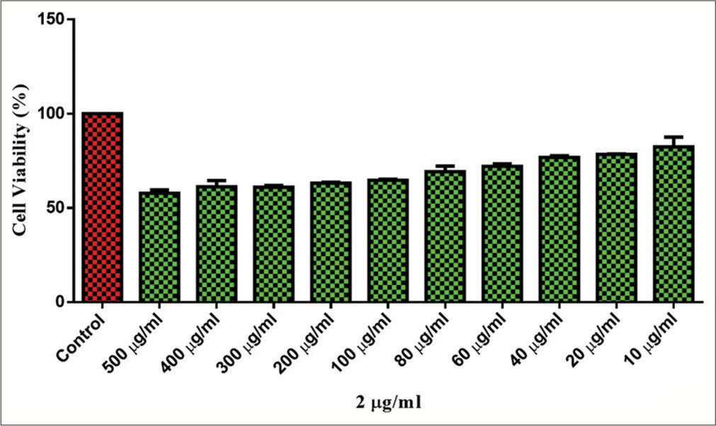
- Cell viability % age of gallstone at different concentrations.
Images of control cells and treated cells
Untreated (controlled) and treated cells at 500, 300, 100, 60, 20, and 10 µg/mL, one can see the lesser distortment signaling cell death, at 500, its more as compare to 300, than to 100, 60, 20, and 10 μg means at 500 µg, it is more toxic [Figure 6].

- (a) Untreated (Controlled) and Treated cells at (b) At 500µg, cell dying stage is maximum (black arrow), (c) At 300, cell death experience (black arrow), (d) At 100, few cells died (black arrow), (e) At 60, cell death commenced (black arrow), (f) At 20, dying activity started (black arrow), (g) At 10, movement of cancer cells started (black arrow).
INTRACELLULAR ROS DETERMINATION/HEP G2
Principle
The assay utilizes the cell-permeable fluorogenic probe dichloro-dihydro-fluorescein diacetate (DCFH-DA), which enters cells and is enzymatically deacetylated into non-fluorescent DCFH. In the presence of ROS, DCFH undergoes rapid oxidation, transforming into highly fluorescent dichloroflurocein (DCF). Fluorescence microscopy captures images at ×20 magnification fields (Life Technology, USA).
Materials Required
IC50 treated cells in the experimental plate, ×1 PBS solution, DCFH-DA (10 mg/mL in DMSO), a fluorescent microscope, and a pipette.
Procedure
For intracellular ROS determination,[18] DCFH-DA staining analysis was employed. MDA-MB-231 cells (1 × 10⁵ cells/well) were seeded in a six-well plate and allowed to mature overnight. The following day, the old medium was replaced with a new medium containing various concentrations of the sample(s) and incubated for 24 h. Subsequently, the plate underwent DCFH-DA[19] staining for 30 min in the dark. Fluorescence staining analysis was performed using fluorescence microscopy (Floid imaging station, Life Technologies, USA). The images were captured with a ×20 magnification lens, and the scale bar used was 125 µm.
Interpretation
ROS molecules play a crucial role in cellular mechanisms and are pivotal in cellular apoptosis. Apoptosis, governed by extrinsic and intrinsic pathways, is influenced by ROS,[18] which are highly reactive and short-lived molecules. Low ROS doses activate cell survival pathways unfolded protein response, nuclear factor erythroidf 2-related factor 2 (UPR, Nrf2), while high doses activate cell death pathways (apoptosis and necroptosis). ROS trigger apoptosis through mitochondrial, death receptor, and estrogen enhances receptor (ER) pathways. Our results indicate that all tested samples effectively induce ROS accumulation in the cell cytoplasm, leading to cell death in the breast cancer cell line (MDA-MB-231). The data suggest that the target samples exhibit a significant capability to induce cell death.
ROS antioxidant analysis-untreated and with treatment, one can see with ROS, there is more bright green signaling cancer cell death [Figure 7].

- ROS Antioxidant analysis -Untreated and with treatment. Green refers to cancer free area while dark spots meant remaining liver cancer cells (black arrow). ROS: Reactive oxygen species.
Bar chart of ROS representing untreated and treated cells represents the treatment given to cancer cells using liquid formulation of gallstone [Figure 8].

- Bar chart of ROS representing untreated and treated cells. Black arrow indicate treatment after ROS - Value to be 1.3 to 1.4. ROS: Reactive oxygen species.
Discussion
The gallbladder stone obtained from surgical removal from a patient era underwent comprehensive analysis,[8-10] shedding light on their elemental composition, microscopic structure, and potential medicinal properties.
Elemental composition
Field emission SEM (FESEM) and EDXRF spectroscopy revealed the presence of essential elements such as oxygen, calcium, silicon, magnesium, and various trace metals in the fossilized Spinosaurus teeth. This elemental profile suggests a complex composition that could contribute to unique properties and potential applications.
Microscopic structure
Examination under a binocular microscope using EuromaxAlpha software unveiled intricate variations and structures within the gallstone. FESEM imaging exhibited a distinctive thread-like pattern, reminiscent of small feathers. This unique microscopic structure adds to the overall intrigue of the stone, indicating potential adaptations during the patient’s lifetime.
Anti-cancer properties
The presence of major elements associated with anti-cancer properties provides compelling evidence of the therapeutic potential of the gallbladder stone. To substantiate this, MTT assays conducted on Hep G2/human breast cancer cells demonstrated significant toxicity. The IC50 value falling below 75 further accentuates the anti-cancer efficacy of the gallstone.
ROS antioxidant test
In the validation phase, a ROS antioxidant test was performed, revealing a fluorescence intensity of 1 with a distinctive green hue. This fluorescence pattern signifies the antioxidant potential of the stone, suggesting its ability to counteract ROS. The indication of anti-cancer and antioxidant properties positions the Spinosaurus teeth as a potential candidate for further exploration in cancer treatment.
Nano chemo drug potential
Given the observed properties, there is potential for the surgically removed gallbladder stone to be utilized as a nano chemo drug in the treatment of breast cancer. The combination of anti-cancer efficacy and antioxidant capabilities opens avenues for innovative therapeutic interventions.
Preservation and future exploration
Considering the need to preserve gallstones, a forward-looking approach involves planting Ayurvedic herbs over the stones using fertile soil. This conservation strategy aligns with the principles of sustainable practices, blending the ancient with the contemporary for holistic preservation.
DISCUSSION
We could see from the tables that although elements such as mercury, gold, zinc, chromium, calcium, chlorine, silicon, aluminum, sodium, oxygen, and carbon are present in all the samples, these gallstones carry carbon and oxygen in maximum concentration. It came to know after a short interview with the patients whose gallbladder stones were examined using SEM energy-dispersive X-ray analysis[20] that they were chain smokers during their youth but adopted a simple life after crossing the age of 38. Although surgeries became possible at the age of 76, stones were examined thereafter.
CONCLUSION
The gallbladder stone presents a captivating intersection of geo-biotechnological and medical science, with the promise of contributing to cancer research and treatment. Further, interdisciplinary studies and ethical preservation practices will unravel additional layers of knowledge encapsulated in these ancient relics.
Acknowledgment
We extend our sincere appreciation to Mr. RP Sharma, for he consented to take his removed gallbladder stone for this invaluable contribution to our research endeavor.
Ethical approval
Institutional Review Board approval is not required because the whole study deals with in-vitro procedures.
Declaration of patient consent
Patient’s consent not required as there are no patients in this study.
Conflicts of interest
There are no conflicts of interest.
Use of artificial intelligence (AI)-assisted technology for manuscript preparation
The authors confirm that there was no use of artificial intelligence (AI)-assisted technology for assisting in the writing or editing of the manuscript and no images were manipulated using AI.
Financial support and sponsorship
Nil.
References
- Ultrasound and CT evaluation of emergent gallbladder pathology. Radiol Clin North Am. 2003;41:1203-16.
- [Google Scholar]
- The gallbladder and bile ducts In: Rumack CM, Wilson SR, Charboneau JW, eds. Diagnostic ultrasound. Vol 1. St. Louis: Mosby-Year Book; 1998. p. :175-223.
- [Google Scholar]
- Imaging benign and malignant disease of the gallbladder. Radiol Clin North Am. 2002;40:1307-23.
- [Google Scholar]
- How should polypoid lesions of the gallbladder be treated in the era of laparoscopic cholecystectomy? Surgery. 1995;117:481-7.
- [Google Scholar]
- The gallbladder and bile ducts In: Rumack CM, Wilson SR, Charboneau JW, eds. Diagnostic ultrasound. Vol 1. St. Louis: Mosby-Year Book; 1991.
- [Google Scholar]
- Does it matter who does ultrasound examination of the GB? Br Med J. 1985;291:389-90.
- [Google Scholar]
- Choledocholithiasis and cystic duct obstruction: Difficult ultrasonographic diagnosis. Radiology. 1983;146:475-9.
- [Google Scholar]
- Jaundice In: Eisenberg RL, ed. Diagnostic imaging: An algorithmic approach. Philadelphia, PA: JB Lippincott; 1988.
- [Google Scholar]
- Rapid colorimetric assay for cellular growth and survival: application to proliferation and cytotoxicity assays. In: J Immunol Methods. Vol 65. 1983. p. :55-63.
- [Google Scholar]
- A critical assessment of the use of microculture tetrazolium assays to measure cell growth and function. Growth Regul. 1995;5:69-84.
- [Google Scholar]
- Detection of total reactive oxygen species in adherent cells by 2’,7’-dichlorodihydrofluorescein diacetate staining. J Vis Exp. 2020;160
- [CrossRef] [Google Scholar]
- Dichloro-dihydro-fluorescein diacetate (DCFH-DA) assay: A quantitative method for oxidative stress assessment of nanoparticle-treated cells. Toxicol In Vitro. 2013;27:954-63.
- [Google Scholar]
- UK institute of physics electron microscopy and analysis group (MAG) Available from: https://groups.iop.org/EM [Last accessed on 2024 May 16]
- [Google Scholar]
- Museum of science-how SEM works. Available from: https://www.mos.org/sln/sem/intro.html [Last accessed on 2024 May 16]
- [Google Scholar]


